

AI & ML Projects to Improve the Cure Rate of Cancer
Our projects focused on improving cancer cure rates and treatment outcomes through innovative AI and ML approaches.
1. Generalizable Quantitative Imaging and Machine Learning Signatures in Glioblastoma
The current state of magnetic resonance imaging (MRI) methods in neurooncology offers excellent potential for providing rich characterizations of brain tumor structural, physiological, and metabolic characteristics, especially in brain gliomas, which are complex and highly heterogeneous cancers.
The current project proposes to refine and translate quantitative imaging methods for glioblastoma (GB) into clinical trials, leveraging machine learning, which aims to help transcend some of these limitations. Our emphasis is on:
- Obtaining rich imaging phenotypes relying on multi-parametric signals, textures, shape properties, spatial patterns derived from atlas registration, and biophysical models of tumor growth
- Integrating such imaging signatures using ML into predictors of clinical outcome, as well as of early recurrence from peri-tumoral infiltration
Our group is part of the Respond consortium NCI R01 grant. We contribute MRI and patient data to the consortium. We subcontract to lead the investigation using AI/radiomics techniques to evaluate GBM patient characteristics of long-term survival and prediction of treatment response to re-irradiation. Besides GB, we also utilized AI/radiomics techniques to study other CNS diseases, including meningioma.
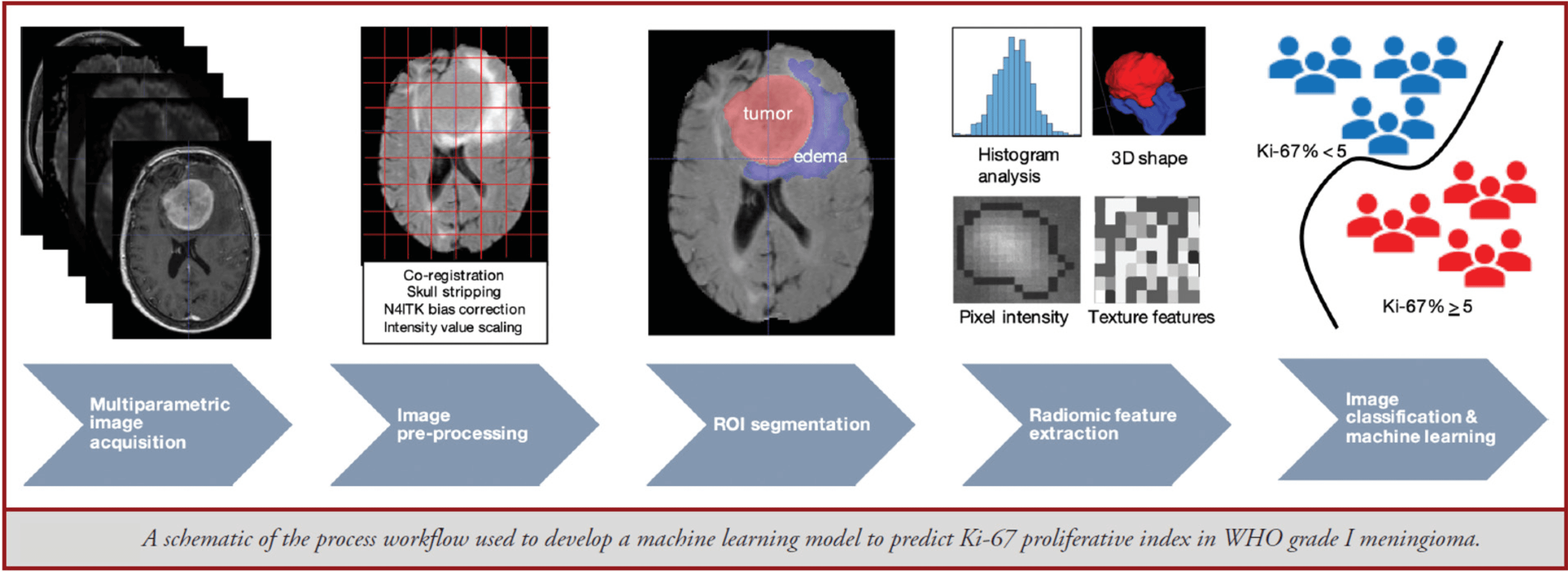
Schematic of the process workflow used to develop a machine learning model to predict Ki-67 proliferative index in WHO grade I meningioma.
2. Ransomware Attack Resiliency in Radiation Oncology using AI
Radiation oncology is susceptible to ransomware attacks because it is the most technology-reliant medical discipline, as the ability to deliver radiation therapy safely relies on multiple complicated software systems.
If a ransomware attack does occur, Radiation Oncology patients are critically impacted as inevitable delays in treatment result in worse disease cure rates and complicated workarounds, resulting in a greater probability of errors occurring.
Jefferson was the most prominent institution impacted by a Radiation Oncology ransomware attack in 2021 that affected more than 40 centers, and we were able to observe firsthand the impact of a ransomware attack in Radiation Oncology.
Based on our ransomware attack experience, our group has developed AI and ML-based methods to help manage patients in a ransomware attack scenario. The developed AI methods can reduce delays in treatment in a ransomware attack setting, resulting in improved cure rates. The project has been awarded a $2,000,000 grant from the Agency for Healthcare Research and Quality. The AI work developed in the grant can reduce patient treatment delays (and therefore improve cure rates), improve safety, and improve outcomes for patients with social determinants of health.
3. AI for Clinical Decision Support (Computational Plasma Proteomics)
Immune checkpoint inhibitors (ICIs) have revolutionized the cancer therapy landscape due to long-term benefits in patients with advanced metastatic disease. However, robust predictive biomarkers for response are still lacking, and treatment resistance is not fully understood.
Current guidelines for the management of metastatic non-small cell lung cancer (NSCLC) without driver mutations recommend checkpoint immunotherapy with PD-1/PD-L1 inhibitors, either alone or in combination with chemotherapy. This approach must account for individual patient variability and host immune factors, often resulting in less-than-ideal outcomes.
To address the limitations of the current guidelines, we developed and subsequently blindly validated a machine-learning algorithm using pretreatment plasma proteomic profiles for personalized treatment decisions. In collaboration with a computational biology company (OncoHost), we developed and subsequently blindly validated a machine-learning algorithm employing pretreatment plasma proteomic profiles for personalized treatment decisions.
Our test demonstrates a strong association between model output and clinical benefit from PD-1/PD-L1 inhibitor-based treatments, evidenced by high concordance between predicted and observed clinical benefit.
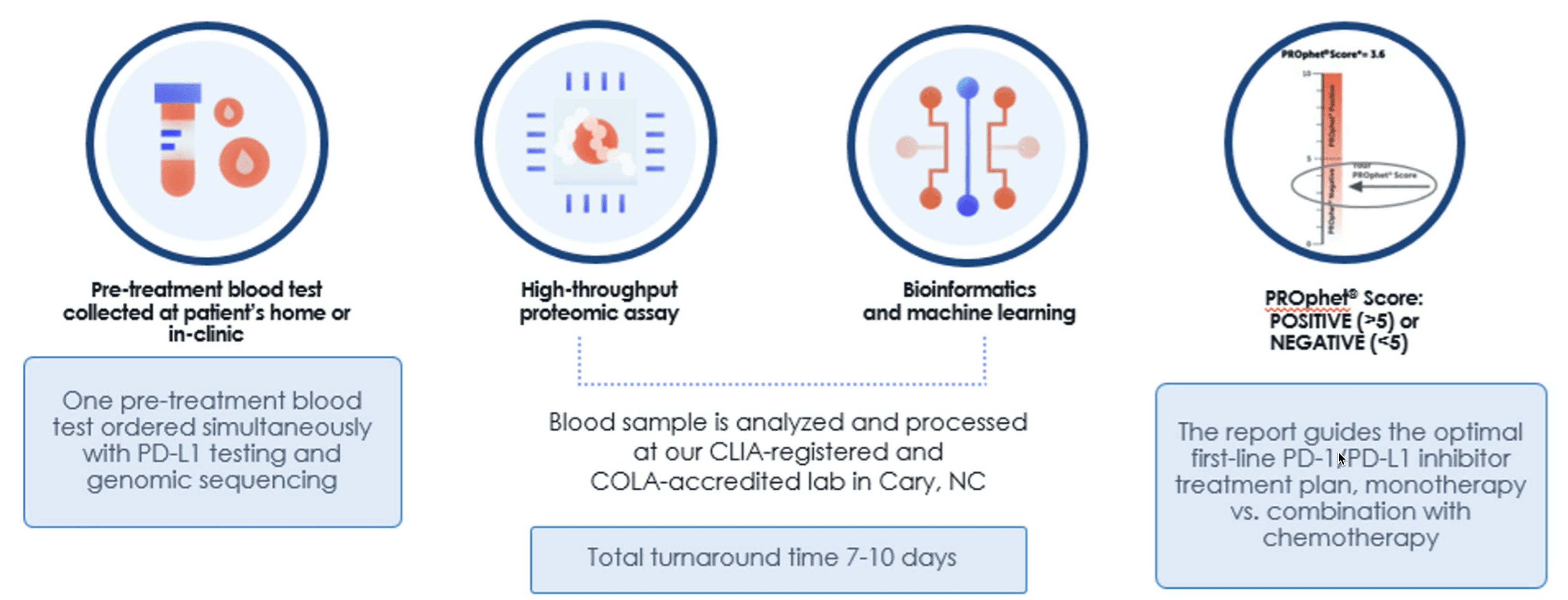
Workflow for the PROphet® test: Blood samples are analyzed using high-throughput proteomics and machine learning to guide optimal first-line PD-1/PD-L1 inhibitor treatment plans.
4. Using AI for Early Detection of Small Brain Metastasis
Stereotactic radiosurgery is being used to treat small brain metastasis. Due to the development of radiotherapy treatment planning and delivery technologies, clinicians are comfortable treating more brain metastases per episode of care.
The challenge is that many lesions are too small to be picked up by radiologists and radiation oncologists. Our group is validating an AI-based software that produces automatic detection of small brain metastasis.
The software has been shown to identify lesions that were missed by human observers. AI-based detection has the potential to provide early identification of small brain lesions and thereby improve the chances of cure.
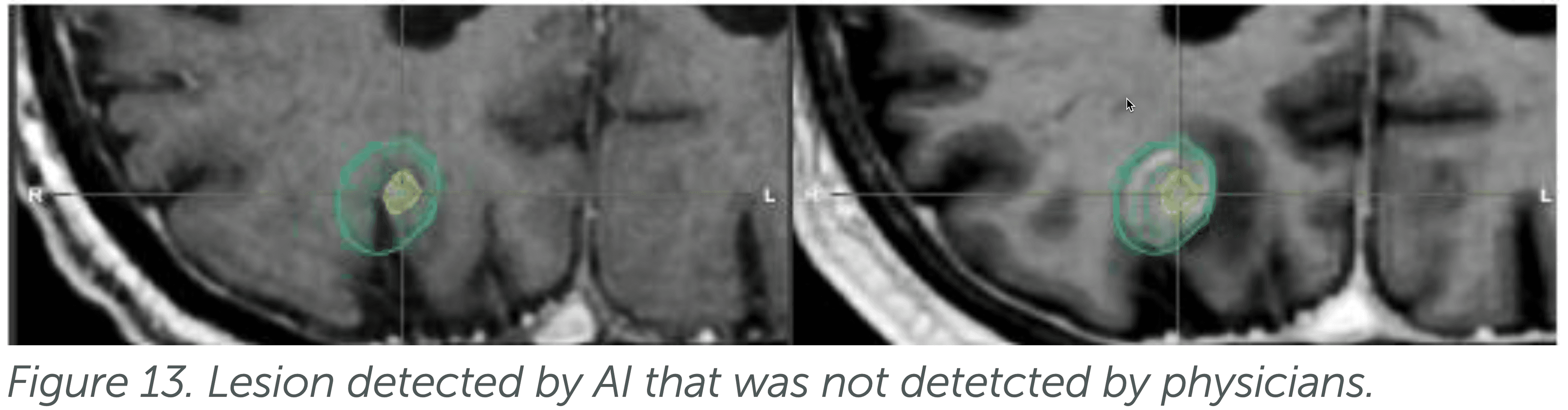
Figure 13: Small brain lesion detected by AI that was not detected by physicians during standard clinical review.
5. AI and ML: K08 Training Grant
A junior faculty, Dr. Lydia Wilson, has submitted a K08 training grant focused on AI titled, 'Novel Functional Imaging to Predict Surgical Side Effects for Lung Cancer Patients.' The proposal aims for Dr. Wilson to become an independent investigator and leader in developing the next generation of AI-based treatment-effect evaluation tools.
The proposed research aims to develop novel imaging and computational tools to improve lung cancer patients' evaluation for resection procedures. Lung cancer is the second-most diagnosed cancer worldwide, and surgical resection is the primary form of definitive treatment. However, lung cancer patients with poor lung function before surgery can develop severe and life-threatening thoracic complications after resection.
The research proposes three cutting-edge computational tools to alter the surgical evaluation paradigm: novel lung-function imaging, voxel-based analysis, and ML prediction. The central hypothesis is that the novel, image-based lung-function evaluation methods can improve the accuracy of post-surgical lung-function prediction from 63% with existing methods to ≥85% using the developed techniques.
The proposed research will test the central hypothesis via two specific aims that will: 1) assess the accuracy of novel lung-function imaging methods in predicting global post-surgical lung function and toxicity measures and 2) develop novel image-processing and machine-learning methods to analyze and predict complex changes in local lung function after resection procedures. The results of this work will enhance the clinician's ability to evaluate patient-specific, pre-treatment thoracic states, improve post-surgical outcome prediction, and provide critical data to guide the next-phase clinical trial.
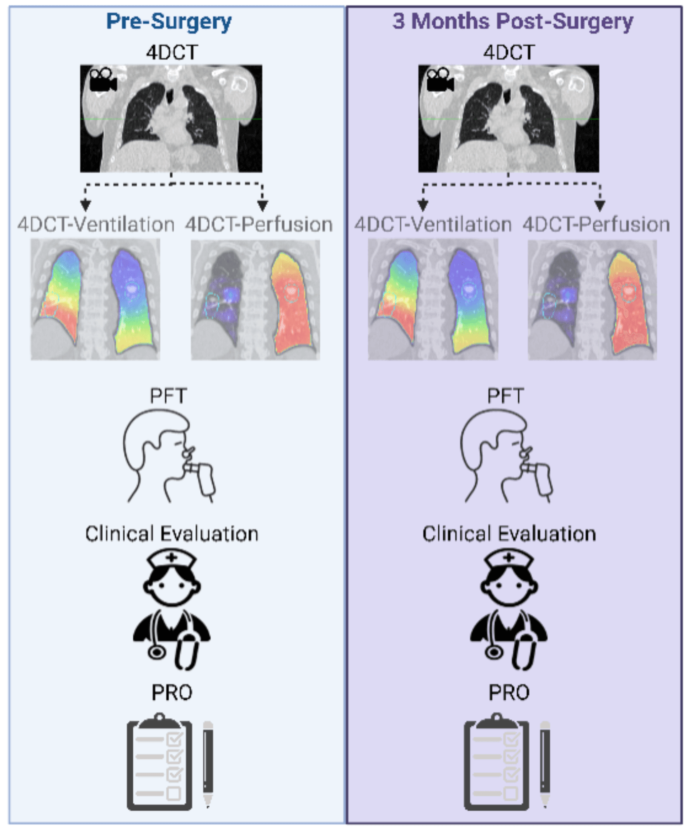
Comparison of pre-surgery and 3-months post-surgery functional lung imaging using 4DCT-derived ventilation and perfusion maps, along with pulmonary function tests (PFT), clinical evaluation, and patient-reported outcomes (PRO).