

AI & ML Projects to Reduce Side Effects
Our projects focused on reducing toxicity and side effects from cancer treatments through innovative AI and ML approaches.
1. AI-based Functional Imaging for Toxicity Reduction
Based on NCI-funded work, department researchers have developed an AI algorithm to repurpose standard-of-care chest computed tomography (CT) imaging to produce functional lung images.
Functional imaging has been incorporated into the radiation treatment planning paradigm for lung cancer patients to reduce toxicity. The AI, imaging-based technology, has been evaluated in a phase II clinical trial (NCT02528942) led by Jefferson investigators, with trial data showing reduced toxicity by 50% compared to standard thoracic radiation.
The AI-based functional imaging technology developed at Jefferson was recently awarded a "Best of Physics" abstract (given to the top 1% of scoring abstracts) at the 2024 national American Society of Therapeutic Radiation Oncology meeting and is being proposed as an NRG cooperative group clinical trial.
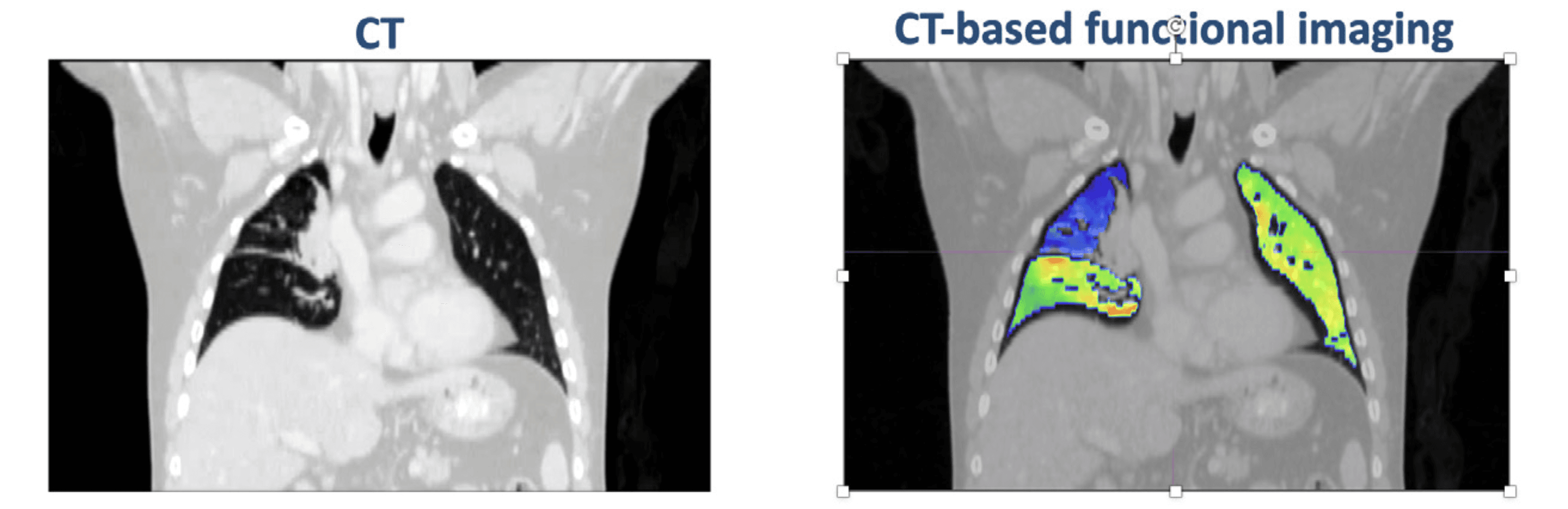
AI-based functional imaging to reduce toxicity from cancer treatment for lung cancer patients
2. Machine Learning to Reduce Toxicity for Prostate Cancer Patients
Jefferson faculty are leading Department of Defense (DOD) work using machine learning to predict and mitigate patient-reported outcome toxicity for prostate cancer patients.
The evaluation of radiation treatment plans for prostate patients relies on singular dose metrics and does not consider the 3D spatial dose distribution. Furthermore, the prostate cancer toxicity paradigm is based on physician-reported outcomes and does not consider the voice of the patient.
This DOD-funded work innovatively solves both challenges using ML methods to determine 3D dose distribution parameters that predict patient-reported side effects. The work has great potential to guide improved treatment planning for prostate cancer patients, which will, in turn, reduce the treatment side effects as reported by the patients themselves.
3. Machine Learning to Reduce Toxicity for Bladder Cancer Patients
Bladder-preserving treatment with radiation is rapidly gaining popularity as an alternative to radical cystectomy to treat muscle-invasive bladder cancer (MIBC) to avoid the physical and psychological impact of the removal of the bladder.
However, current radiation treatment planning for MIBC is based on a patchwork of older trials using antiquated radiation schedules and dosimetric constraints. Consequently, the short- and long-term urinary, bowel, and sexual toxicities of radiation for MIBC can be considerable.
Patients can live for many years after MIBC treatment; preventing short- and long-term toxicities and optimizing quality of life is essential (QoL).
The aims are to describe rates of real-world, patient-reported toxicities and to develop and validate deep learning models incorporating demographic, clinical, behavioral, dosiomic, and radiomic data for actual multi-endpoint prediction of patient-reported toxicities among MIBC patients treated with radiation.
4. AI and Standard of Care PET Imaging to Predict Cardiotoxicity
Twenty percent of patients receiving radiation to the chest experience clinically significant cardiac toxicity. Models for predicting which patients are at higher risk of developing cardiac toxicity are population-based and lacking in accuracy and robustness.
Funded by SKCCC seed grants, Jefferson Radiation Oncology researchers have developed an AI algorithm to repurpose standard-of-care staging PET scans to extract clinically significant cardiac information to improve the prediction of cardiotoxicity.
The AI algorithm has been filed as a provisional patent, is being evaluated in an investigator-initiated clinical trial, and has been scored as an NCI R01 grant with a re-submission planned.
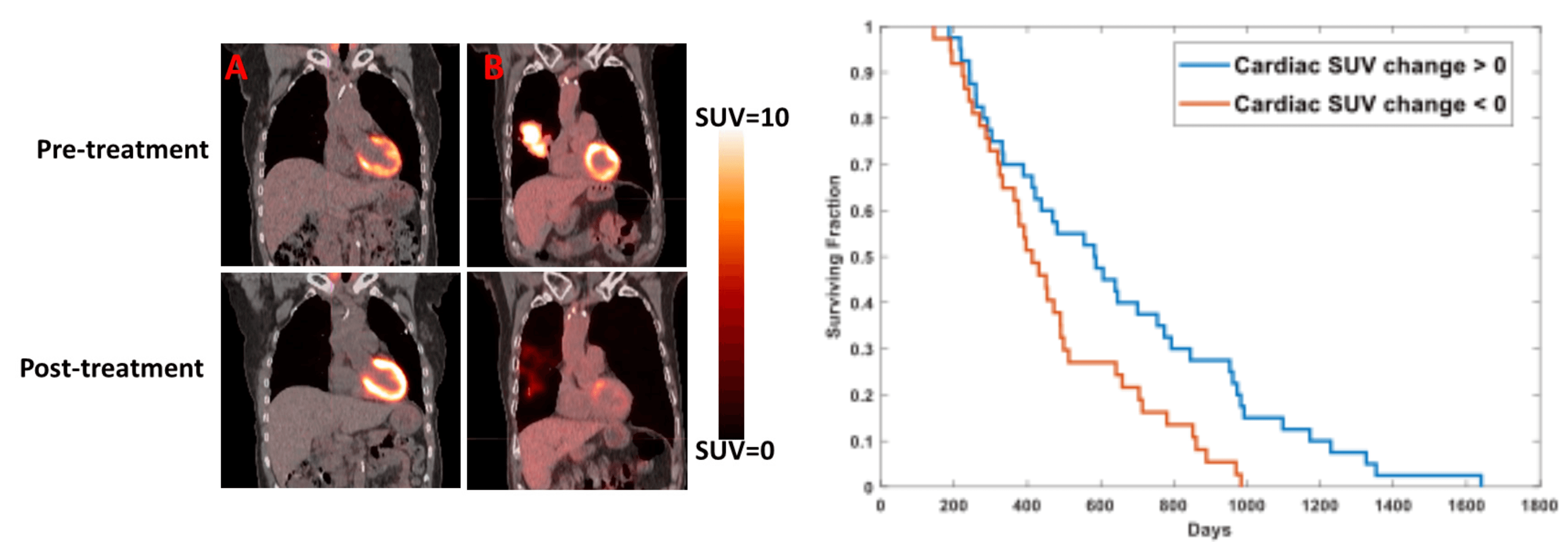
Left: PET/CT scans showing pre- and post-treatment cardiac uptake. Right: Survival curves based on cardiac SUV changes.
5. AI-based Dose Prediction from Diagnostic Imaging
Multi-disciplinary clinics are the standard of care in NCI-designated cancer centers. When Radiation Oncologists meet with patients in a multi-disciplinary clinic, it would be clinically beneficial to know how much radiation dose will be delivered to critical organs to provide better-informed guidance to patients.
However, knowledge about the dose distribution cannot be known when physicians meet with patients in a multi-disciplinary clinic because the radiation treatment planning process typically takes two weeks.
We have developed a patent-pending AI algorithm to predict the radiation dose distribution from diagnostic imaging (available during the multi-disciplinary clinic). Having the radiation dose information at the time of a multi-disciplinary clinic will enable clinicians to make better-informed decisions about multi-disciplinary care.
6. Radiotheranostics Dosimetry Using AI
Radiotheranostics is a form of radiotherapy where alpha or beta particles emitted from injected molecular-targeted radionuclides are used to treat malignancies. Radiotheranostics has been shown to improve survival for men with stage IV prostate cancer significantly and is being investigated as a promising treatment for other types of malignancies.
The challenge with radiotheranostics is that the dosimetry still needs to be discovered. In other words, when the radionuclide is injected, it is unknown how much dose goes to critical organs.
Using funds from a recently awarded Philadelphia Prostate Cancer Biome Project, our team is developing a microdosimetry laboratory that uses a high-resolution alpha/beta camera combined with advanced AI techniques. The microdosimetry lab will develop strategies to determine how much dose is being deposited during radionuclide therapy and enable the development of personalized treatment, thereby reducing the rate of side effects for patients treated with radionuclide therapy.
7. Breast Voxel-based Analysis for Toxicity Prediction
Nine in ten women diagnosed with breast cancer survive five years or more, but most are left with long-lasting side effects of their curative treatments. Our AI-based project proposes leveraging novel voxel-based computational analysis to identify which tissues are most radiosensitive and critical in predicting which patients may get side effects.
The voxel-based analysis comprises two primary steps: 1) spatial normalization, where we map individual patient anatomy to reference anatomy, and 2) statistical inference, where we perform voxel-wise statistical tests to identify spatial associations between medical imaging data and an effect of interest.
The developed methods will identify the tissues most sensitive to radiation and reveal how to treat breast cancer more effectively by avoiding the identified tissues. The results of the study can lead to reduced side effects and improved quality of life for patients diagnosed with breast cancer receiving radiotherapy.
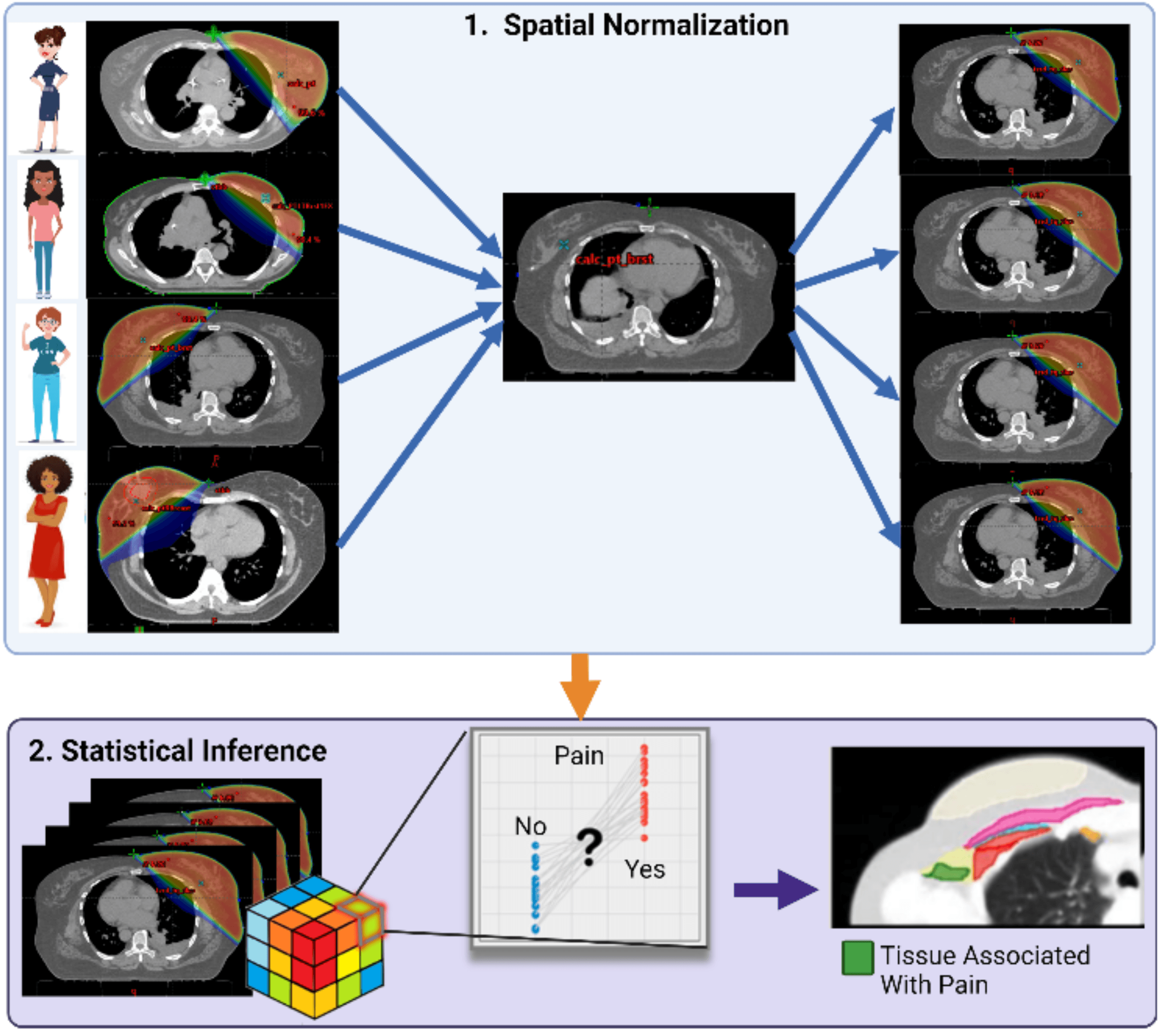
Voxel-based analysis workflow: 1) Spatial normalization maps individual patient anatomy to reference anatomy, 2) Statistical inference identifies tissues associated with radiation-induced pain.